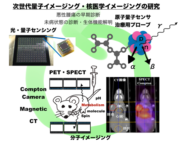
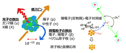
核医学イメージング技術は悪性腫瘍(がん)の早期診断や生体機能や生体内の分子動態の可視化に広く使われています。1原子をラベルとして用いることで非常に高感度なセンシングが可能となります。研究室ではこの様な技術の高精度化に加えて、新たな未来の核医学イメージング技術の研究を行っています。また原子や原子核・光などの量子をもちいて高感度な磁場や電場・化学状態などのセンシングを行う量子センシング技術の研究を行っています。加えて原子核から放出される放射線を高精度に計測し制御する技術の研究も行っています。
研究テーマ
量子イメージング・核医学診断・治療の高度化に関連する研究
Quantum Imaging, Nuclear Medicine, Radio-theranostics
原子核・素粒子・スピンを利用した生体センシングの研究
Biosensing based on spin, atomi nucleus, elementary particles
量子もつれ光子対による原子核ー多分子間相互作用プローブを活用した診断治療学の創生
Creation of novel thranostics using nuclear-multimolecular interaction probes by quantum entangled photon pair
カスケード放出ガンマ光子を用いた診断 多光子を用いた新イメージング手法の研究
Novel Medical Imaging Method based on Multi-Photon cascade gamma-rays
Research on Magnetic field and ionizing radiation
PET(Positron Emission Tomography)の研究 生体機能や分子を可視化する装置の開発
Research on Positron Emission Tomography and molecular imaging
脳機能、生体機能イメージング、意識に関する研究
Functional imaging, brain imaging, consciousness
量子生体センシング ~ 量子技術を生体に応用することで超高感度な生体計測の研究
Highly sensitive biomedical imaging based on quantum feature/ Quantum Biology
環境モニタリングカメラの開発 福島等における環境放射線の可視化技術を開発
Enviomental Radiation Monitoring, Fukushima Disaster Monitoring
量子検出多チャンネルLSIの開発 イメージングに必要な高度な信号処理技術を開発
Electronics development, integrated circuit for radiation imaging sensors
3次元半導体センサの研究開発 SOI技術を用いた3次元電子、光子可視化技術の開発
B/GdNCT( Neutron Capture Therapy)の研究 中性子を用いた新たな治療法の開発
New therapy with Boron/Gd/Rh neutron capture therapy
MSGC・GEMの開発 ガスを用いたX線や中性子の検出技術の研究開発
Development of gaseous detector for X-ray and neutron
薬剤イメージングの高度化 多分子の同時撮像や体内動態の研究
Drug delivery imaging, multi-isotope, multi-molecule imaging
超電導単一光子検出器に関する研究 量子効率の高い光子検出技術開発
Superconducting single photon detectors with high quantum efficiency
内視鏡腹腔鏡型RI検出器の開発 術中で悪性腫瘍をモニタリングする技術の開発
Intraoperative endoscopic/laparoscopic diagnosis device
ミュオンイメージングの研究
Muography, Muon imaging detectors
多光子技術を用いた医用量子イメージングの開発
Quantum medical imaging with multi-photons
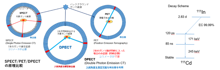
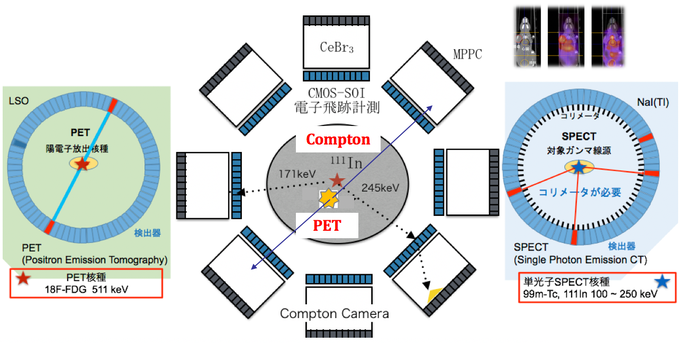
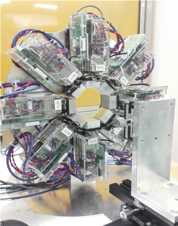
従来の核医学診断法であるPETでは解像度、SPECTでは撮像感度の点で原理的限界があります。我々は複数ガンマ線を同時に放出するカスカード放出核種では時間相関から空間情報が得られる点に着目し、ガンマ線入射方位を特定できる反跳電子追跡型ガンマカメラを駆使した多光子ガンマ線時間/空間相関型断層撮像法を新たに考案しました。本撮像法は複数光子間の相関を用いて体内放射能濃度を高分解能・高感度・高S/N比で描出し、従来のPET、SPECTの本質的な限界を突破する画期的な手法となることが期待されます。本研究では新手法の分解能・感度・S/N比等の諸特性の評価を狙い、象徴的な半球型試験装置を製作し、分子イメージングを革新する計測原理を追求する。具体的にはIn-111標識ペプチドを用いて2光子放出核種検出1分子イメージングを実現し、日本発の革新的ガンマ線診断技術の確立と、44Scなどの多光子核種への展開を図っています。
In conventional nuclear medicine imaging, PET and SPECT suffer from the resolution and sensitivity originated from the imaging principle. PET and SPECT depends on the tomography principle with Radon transform. We porposed a new imaging principle utilizing multi-photon detection and imaging using angular resolving detectors. This method enables the detection of single molecule as a point and could be the new modalities of molecular imaging.
コンプトンPETハイブリッドカメラの開発
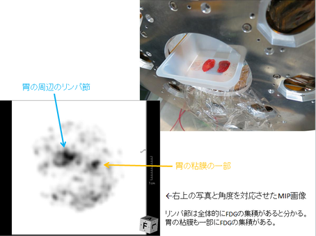
PET (Positron Emission Tomography)はその名の通り電子の反物質である陽電子放出核種を用いて、陽電子−電子対消滅から生じる511 keVのガンマ線を測ることで画像化を行いますが、単一のエネルギーであるため複数の分子の可視化は困難です。一方でSPECT (Single Photon Emission CT)はそれよりも低い数百keV程度のエネルギーをコリメーターを用いて撮像しています。PETとSPECTの分子はこれまで同時撮像が困難でしたが 研究室では新たにコンプトン散乱原理とPET原理に基づく装置を開発し初めて両原理をもちいた同時撮像に世界で初めて成功しました。
PET(Positron Emission Tomography) utilizes annhilation gamma-rays (511 keV) generated by positrons (anti-matter of electrons) with electrons for imaging, but only visualize one tracer. SPECT utilizes single photon emitters using mechanical collimation. There were no method to visualize multi-tracers since it is diffcult to combine two modalities. Our group have developed and demostrated the combination of Compton imaging and PET imaging for the first time by visualizing PET and SPECT tracers in vivo.

高分解能PET(Positron Emission Tomography)の研究
High resolution Positron Emission Tomography
東北大学(吉川先生、鎌田先生)開発の新規高エネルギー分解能シンチレータ Pr:LuAG/Ce:GAGG
東工大(片岡先生:現早稲田大学)開発の144チャネルAPDアレー/ SiPM
東大開発の時間幅方式のASICを組み合わせた純国産のAPD/SiPM-PETシステムを開発しました。
空間分解能でサブmmの解像度を実現しており、企業と共同研究を行っています。
We have developed a sub-milimeter resolution PET scanner in collaboration with Tohoku University (Prof. Yoshikawa, Prof. Kamada), Waseda University (Prof. Kataoka) with newly developed gamma-ray detection crystal, photon sensor array and application specific integrated circuit (ASIC). We are now developing a product.
東京大学医学部と共同にて随時性能評価を行なっています。
高分解能のPETのシステムは生体内の分子イメージングや新規に開発された
薬剤の動態解析などに近年重要な役割を果たしています。
PET/SPECT薬剤と集積イメージング技術の研究
PET/SPECT tracer imaging and molecular imaging
高感度に病変や動態を検出可能な薬剤とそれと組み合わせるイメージング技術の開発を東京大学アイソトープセンター、東京大学薬学部、理化学研究所などの共同で行っています。PETとSPECT薬剤は様々な核種が存在しており、それぞれの特性を生かしたイメージング研究を探索しています。
We are investigating the new combination of tracers and imaging technology by collaborating with UTokyo Isotope Science Center and UTokyo department of Pharmacy and RIKEN. There exist many kinds of radioactive trecers and new imaging principle with different radioisotope is under investigation.
環境放射線モニタリングシステムの開発
Envionmental radiation monitoring system
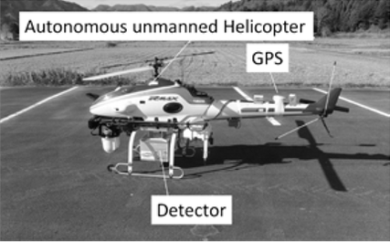
医療用途および環境モニタリング用途としてGAGG結晶およびAPDやdigital SiPMなどの光検出器アレイを用いたコンプトンカメラシステムの開発を行いました。コンプトンカメラシステムはガンマ線が検出器に落とすエネルギーからガンマ線の入射角度を推定する手法で多チャンネルの検出器をもちいることでガンマ分布の可視化が可能になります。特に福島第一原子力発電所事故後、無人ヘリコプターを利用した放射線レベルのモニタリングが重要となっており本研究室ではJAEAや東北大学と共同で開発を行なっています。
We are developing new Compton imaging system based on new scintillation materials and new photon sensors. Compton imaging is useful to detector wide-band multi-energy gamma-rays based on Compton scattering kinematics. We have developed a autonomous unmanned helicopter system with Compton camera to monitor the distribution of cesium 137 in Fukushima area for the improvement of environment.
量子センサ用多チャンネルLSIの開発
Integrated electronics for radiation detectors
PETやコンプトンカメラなどの近年の高精度のイメージングシステムには多チャンネルの信号処理技術が必須となります。マルチチャネル放射線計測用に適したASIC (Application Specific Integrated Circuit)の開発を行なっています。研究室では新しく信号処理方法を考案しADCが不要な多チャンネルシステム (Time over Threhold)を実現しています。
Sophisticated electronics are mandatory for the modern radiation imaging system, such as Compton camera and PET. We are developing multi-channel multi-functional integrated circuit with sub-micron CMOS technology.
またミュンヘン工科大学(TUM)と共同で新型次世代PETの共同開発を行いました。
また放射線医学総合研究所(QST-NIRS)と共同で重粒子線治療下のPET用検出器の開発を行なっています。
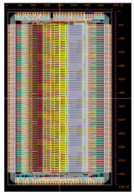
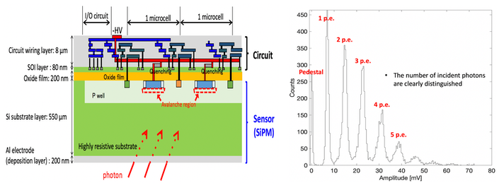
超高感度検出器 SOI-SiPMの開発
Highly sensitive SOI-SiPM
多くの放射線計測システムでは放射線を光に変換するシンチレータ検出器とともに優れた光検出器が必要となります。本研究室ではフルデジタルの最新の光検出器であるDigital SiliconPhotomultiplierの評価をドイツミュンヘン工科大学と共同で行なっています。またDigital SiPMを用いたPETやコンプトンカメラシステムの開発も行なっています。近年シリコン半導体を用いた1光子検出技術が可能となってきており、これらの光センサと回路技術を用いた開発も進めています。可視光1つ1つを見ることが可能な技術は様々な生体応用につながっています。
In many radiation detector system, high grade photon sensor is required combined with conversion materials from radiation to visibule sensors. We are evaluating digital silicon photomultipliers with TUM group and using it for Compton and PET imaging. We are also developing SOI based silicon photomultipliers with a single photon detection capability and integrated circuit. Bottom figure shows single photon detection capability.
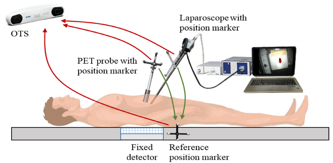
内視鏡・腹腔鏡型PET技術の研究開発
Endoscopic/Laparoscopic PET technology.
通常の核医学診断技術は術前術後に行いますが、実際の手術支援、切除範囲の縮小化には術中の悪性腫瘍部位の診断技術が求められています。研究室では術中に利用可能なPETの可能性を模索し研究開発を行っています。
We are also developing intraoperative PET imaging/diagnosis system which is important to reduce the resection area and increase the quality of life.
上記の他にも量子効果を用いた新たなイメージング技術や原子核の特性に関わる研究など様々な研究を実施しています。